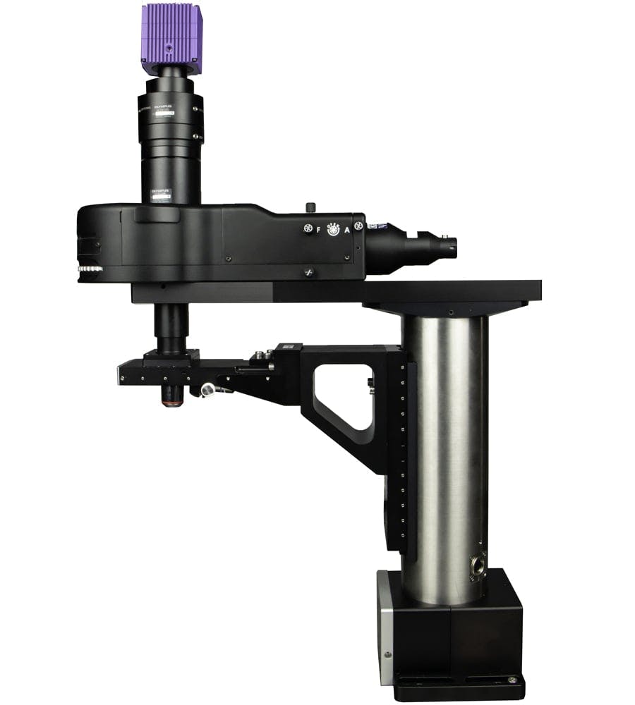
Scientifica VivoScope | Multiphoton Imaging System

Scientifica VivoScope

The VivoScope is a slimline upright microscope. When combined with a multiphoton scan head, it forms the foundation of the finest in vivo imaging system for two-photon behavioural studies.

The VivoScope is ideal for larger in vivo samples, linear or spherical treadmills, large stereotaxic frames or other virtual reality set-ups. The extended position of the scan head gives more room between the light path on your sample and the microscope frame.

Benefits

Extra space

The extended scan head gives more room between the light path onto your sample and the microscope frame without compromising stability (205.94 mm up from 130 mm).

Galvo or Resonant

The VivoScope is available with both galvo and resonant scan head options, depending on your experimental requirements.

Acoustic isolation

Our custom scan head enclosure integrates acoustic insulating foam to dramatically reduce the audible noise created by the resonant motor without affecting performance.

Patch under multiphoton illumination

The fast frame rate provides the visual feedback required to position your patch pipette accurately under infrared laser light.

Deep imaging for large populations of cells

Tailored to large back aperture objectives, the entire field of view can be quickly scanned to monitor the activity from a large number of cells.

The VivoScope microscope frame is available with resonant or galvo scan heads and a variety of detectors including the MDU, MDU XL, ChromoFlex and FLIM Upgrade kit. Take a look at our buyer’s guide to see our full multiphoton range and all scan head, frame and detector options available.

The VivoScope in action at the Allen Institute

The Allen Institute for Brain Science is using three VivoScopes to collect data for the Allen Brain Observatory, an ongoing project to visualise the brain in action. Watch the video below for more information:


Control options

The VivoScope can be fully controlled in Scientifica's own SciScan software. It can also be driven by Vidrio Technologies ScanImage or custom software.

Design & Specifications

Scanning mirror (y-galvo)
3 x 5 mm 8315KL scanning mirror (MicroMax 671HP driver)
Scanning mirror (x-galvo)
2 kHz in bidirectional scan mode (4 fps @ 512x512 px), 1 kHz in unidirectional scanning mode (2 fps @ 512x512 px)
Scanning mirror (x-resonant)
3 x 5 mm 8 kHz CRS series scanner from Cambridge Technologies (15 fps @ 1024 x 1024 px, 30 fps @ 512 x 512 px, 60 fps @ 512 x 256 px)
Beam input height
Beam input height
280.6 mm
Input beam diameter
Input beam diameter
<3 mm
Beam diameter at back aperture
<20.1 mm
Beam expansion
Beam expansion
6.7x
Relay lens expansion
1x
Lens coating
Lens coating
700-1200 nm (Rav <0.5%)
Maximum scan angles (galvo)
+/- 10 optical degrees
Maximum scan angles (scan head)
+/- 7.6 optical degrees
Galvo voltage/optical degree
0.25 V
Turning mirror size
Turning mirror size
45 x 64 x 6 mm
Turning mirror coating
Protected Silver (98% reflectivity)
Typical field of view
~600 µm2 (16x), ~500 µm2 (20x)
Scan control
Scan control
SciScan, ScanImage 5

VivoScope Multiphoton Microscope SchematicVivoScope Multiphoton Microscope Schematic

VivoScope Multiphoton Microscope Schematic

VivoScope moving microscope with a Tiltable Objective Mount (TOM) and 100um Piezo

Erskine, A., Ackels, T., Dasgupta, D., Fukunaga, I., Schaefer, A. (2019). Mammalian olfaction is a high temporal bandwidth sense. bioRxiv https://www.biorxiv.org/content/biorxiv/early/2019/03/09/570689.full.pdf

Roland, B., Deneux, T., Franks, K., Bathellier, B. and Fleischmann, A. (2018). Odor identity coding by distributed ensembles of neurons in the mouse olfactory cortex. eLIFE http://dx.doi.org/10.7554/eLif...

Sun, B., Wang, M., Hoerder-Suabedissen, A., Xu, C., Packer, A., & Szele, F. (2022). Intravital Imaging of the Murine Subventricular Zone with Three Photon Microscopy. Cerebral Cortex. https://doi.org/10.1093/cercor/bhab400

Sun, L., Chen, R., Bai, Y., Li, J., Wu, Q., Shen, Q., Wang, X. (2018). Morphological and Physiological Characteristics of Ebf2-EGFP-Expressing Cajal-Retzius Cells in Developing Mouse Neocortex. Cerebral Cortex https://doi.org/10.1093/cercor/bhy265

Garrett, M., Manavi, S., Roll, K., Ollerenshaw, D., Groblewski, P., Ponvert, N., Kiggins, J., Casal, L., Mace, K., Williford, A., Leon, A., Jia, X., Ledochowitsch, P., Buice, M., Wakeman, W., Mihalas, S. and Olsen, S. (2020). Experience shapes activity dynamics and stimulus coding of VIP inhibitory cells. eLife, https://elifesciences.org/articles/50340

Umpierre, A., Bystrom, L., Ying, Y., Liu, Y., Worrell, G. and Wu, LJ. (2020). Microglial calcium signaling is attuned to neuronal activity in awake mice. eLife, https://elifesciences.org/articles/56502

饰品

Movable Periscope Bracket (MP-4010-30)

This bracket attaches the periscope directly to the XY stage allowing the customer to move the microscope in X and Y whilst maintaining perfect laser alignment.

Contact Form

Contact us

*为必须填写内容

选择感兴趣领域