
Scientifica VistaScope
样品数据
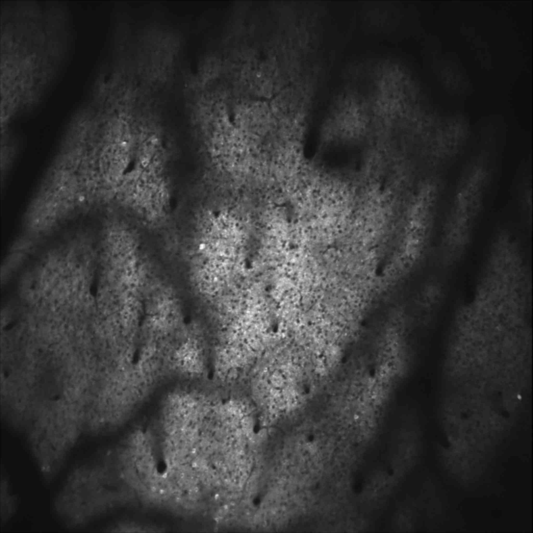
The image shows a 1.8mm square region of mouse somatosensory cortex, layer II/III expressing GCaMP6s.

Over the industry leading FOV of 1.8mm (with a 16x objective), the VistaScope has less than 10% variance in fluorescence intensity (typical: < 5% drop to the edges)
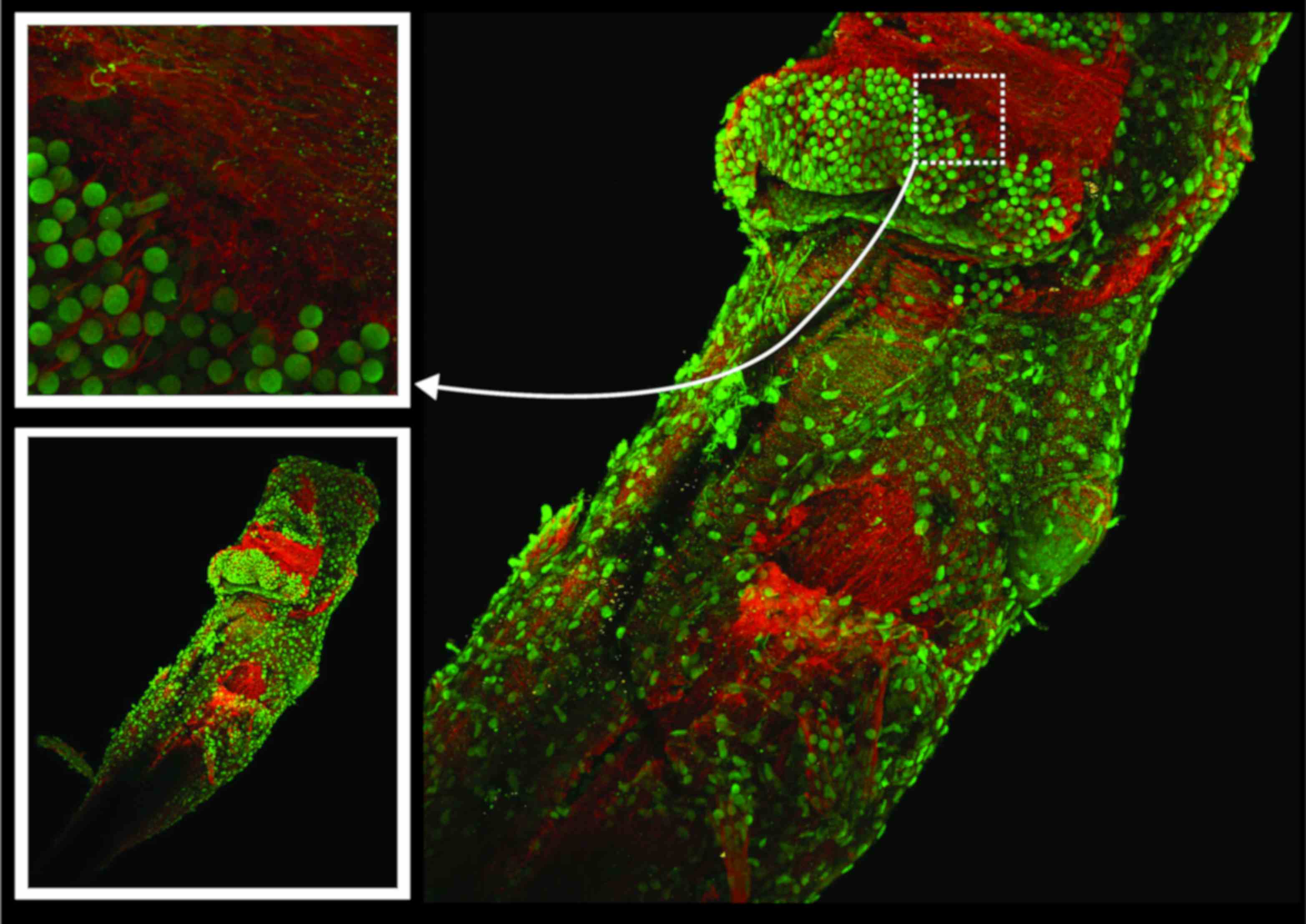
Image captured at the 2023 CSHL imaging courses using the VistaScope: Brain and brain stem of Xenopus laevis stained for tubelin and nestin.
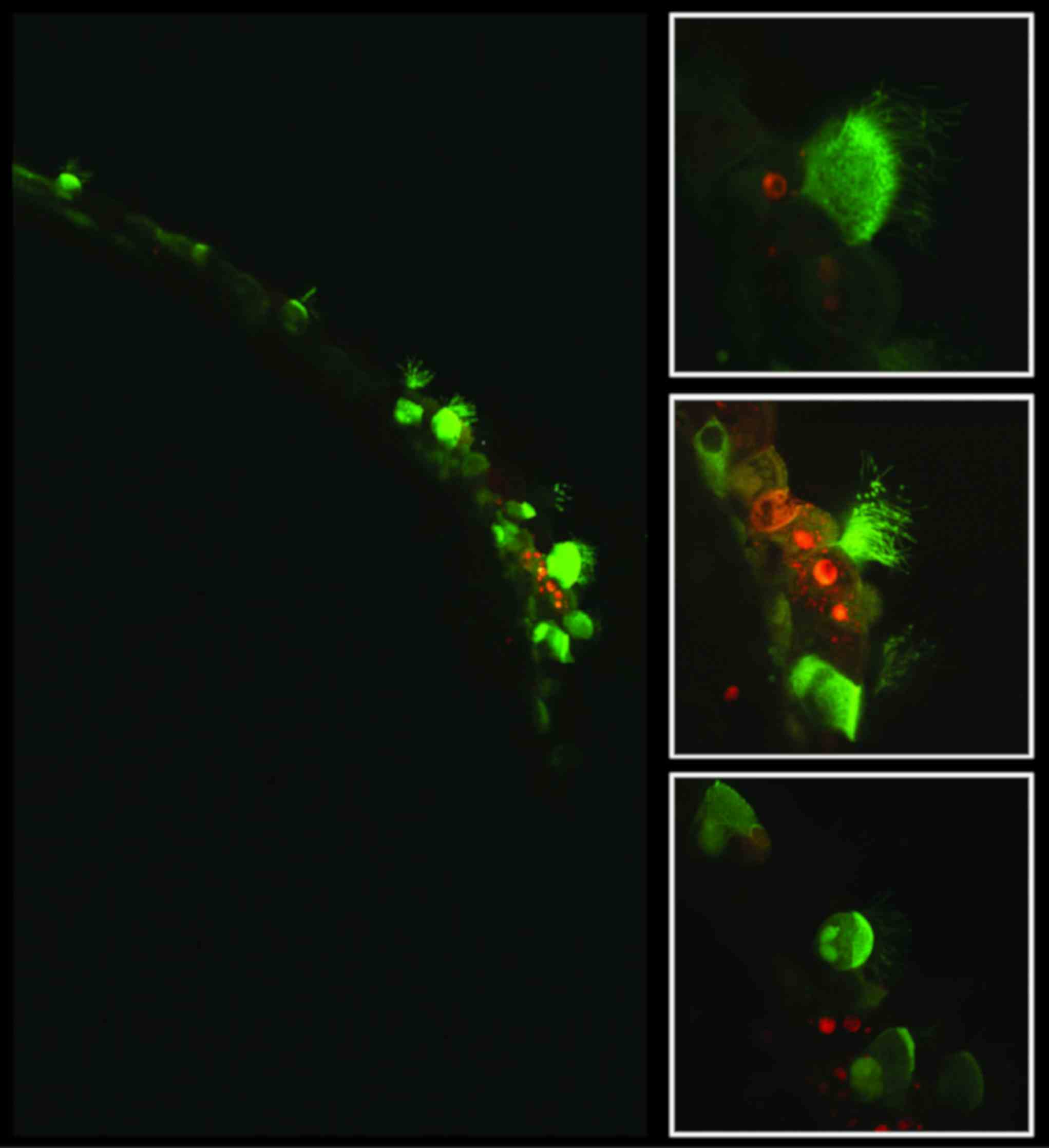
Image captured at the 2023 CSHL imaging courses using the VistaScope: Multicileated cells inside Xenopus.
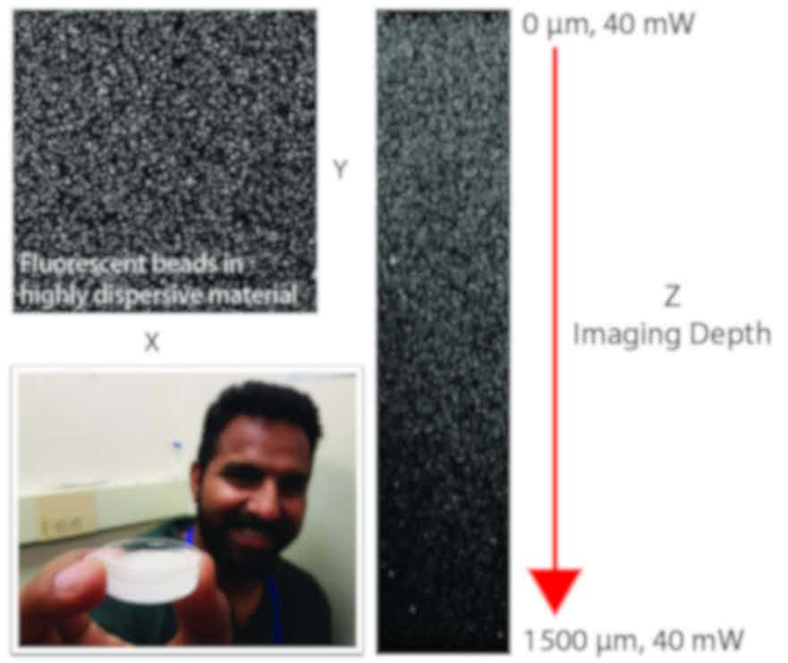
Image captured at the 2023 CSHL imaging courses using the VistaScope: Imaging fluorescent green beads deep with a dispersive sample.
标准系统
We can customise this system to suit your unique experimental needs. Please contact us to discuss your requirements.
SliceScope or VivoScope
Choose from the MDU or MDU XL
Configured as GG, RG or RGG scan paths
Imaging
- Field Number: FN40
- Resonant imaging: 30fps imaging at 512x512 across a 1.18mm FOV
- Wavelength range: 700 - 1700 nm
- Fluorescence homogeneity: <10% intensity roll-off over full width